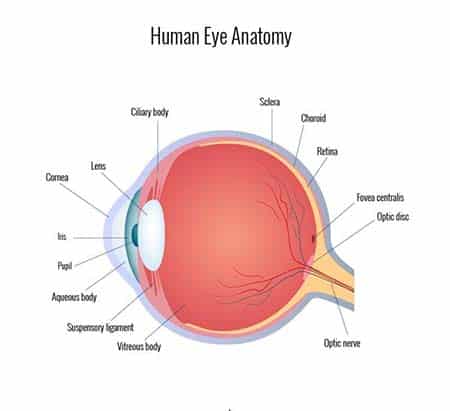
Human Eye Parts and Terminologies
 Most of us take our eyes for granted, so have you ever truly examined the anatomy of an eye? It’s only when something is not right that we even think about them, for the most part. They seem such a small part of our anatomy, but did you know that the eye is one of the most complex organs in the body? The anatomy of the human eyes is not really surprising if you consider the complicated task the eyes handle – providing vision.
Some parts of the eye are easily seen, just by looking at it, though we might not know what we’re seeing.
- The iris and pupil– The iris, the colored part of your eye, a ring-shaped tissue that controls the amount of light that enters the eye. The pupil is the opening in the middle of the iris. The iris has a ring of muscles that cause the pupil to grow smaller in bright light or become larger to let in more light when it’s dim or dark.
- The sclera– The sclera, the white part of your eye, is actually a leather-like tissue that surrounds the eye and gives it shape.
- Conjunctiva– The conjunctiva is a thin, clear skin that covers the front of the eye, including the sclera and the inside of the eyelids. It helps to keep bacteria and other foreign material from getting behind the eye.
- Cornea– The cornea is a clear layer at the front and center of the eye-so clear that you may not even realize it is there. It’s located just in front of the iris and helps focus light as it enters the eye. Contact lenses rest on your corneas.
A closer examination of the eye reveals a very detailed structure, with many different components.
- Extraocular Muscles– Six extraocular muscles are attached to each eye to help move the eye left and right, up and down, and diagonally.
- Crystalline lens– The lens is a clear, flexible structure located just behind the iris and the pupil. It is surrounded by a ring of muscular tissue called the ciliary body, and these two components work together to control the focus of light passing through the eye.
- Optic Nerve– The optic nerve is actually 1 million nerve fibers that transmit nerve signals from the eye to the brain. The front surface of the optic nerve, visible on the retina, is called the optic disk.
- Vitreous Cavity-The vitreous cavity, behind the lens and in front of the retina, is filled with a gel-like fluid, called the vitreous humor, which helps maintain the shape of the eye.
- Retina– The retina converts light signals into nerve signals. Then these signals are sent to the optic nerve, which carries the signals to the brain, which processes the image. The retina contains rods, which work in low light, but don’t allow you to see color, and cones, which allow color, but require more light.
- Macula– The macula is in the central part of the retina, and is responsible for giving you sharp central vision.
All of these individual parts of the eye work together to gather and focus light, to transform it into a picture we can interpret. If you really understand what’s involved in sight, you’ll understand what a remarkable organ it is — it’s one of the reasons Dallas LASIK surgeon Dr. Tylock is so passionate about his work! Such complex machinery deserves special care to keep it working at top capacity.


